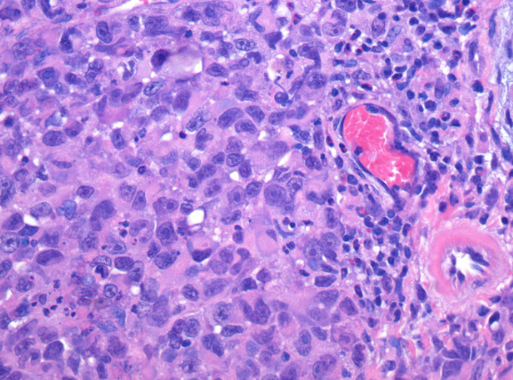
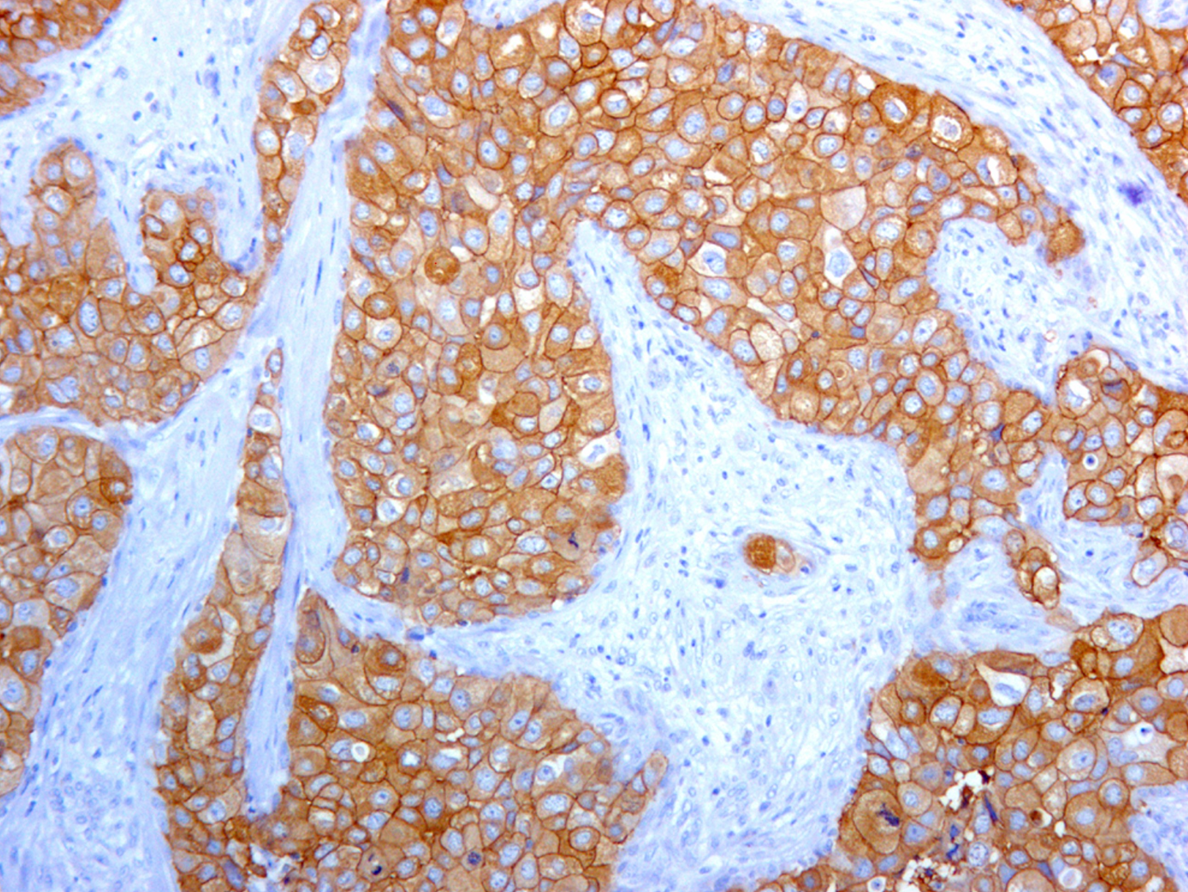
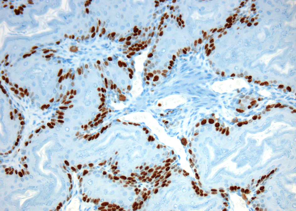
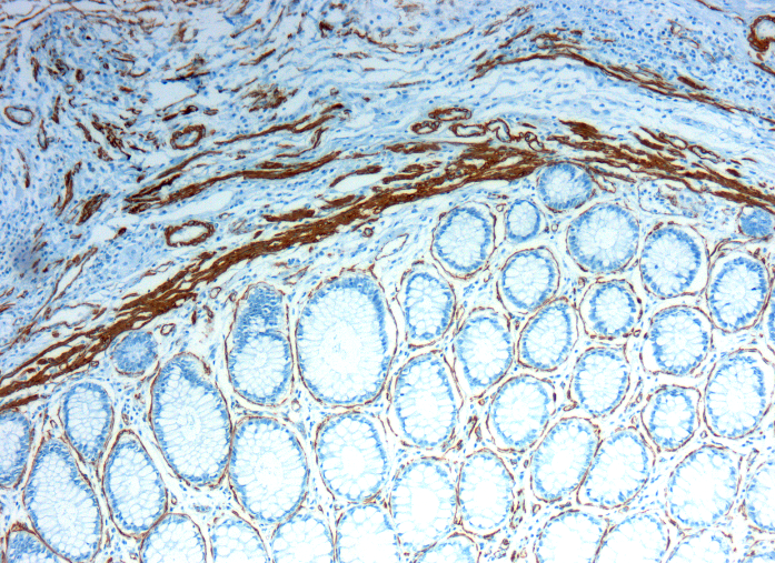
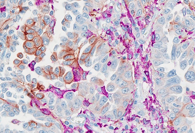
The Research Pathology Facility (RPF) is a full-service research facility which is part of the Montreal Center for Experimental Therapeutics in Cancer (MCETC) and has been established to provide services for investigators associated with both the MCETC and the Lady Davis Institute for Medical Research (LDI), as well as other universities and companies. Our objective is to provide pathological and histological services to support research at the LDI. The mandate of the RPF is to accelerate translational research at the MCETC.
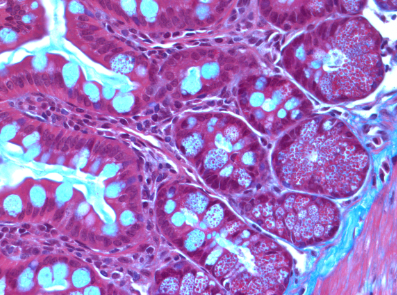
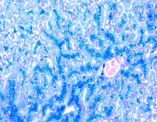
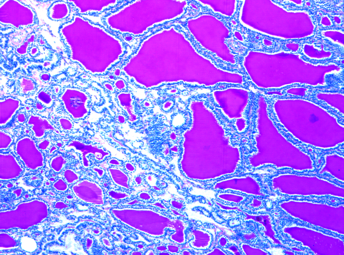
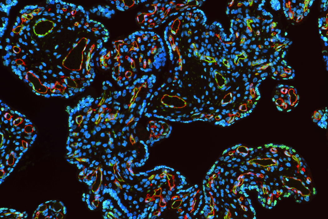
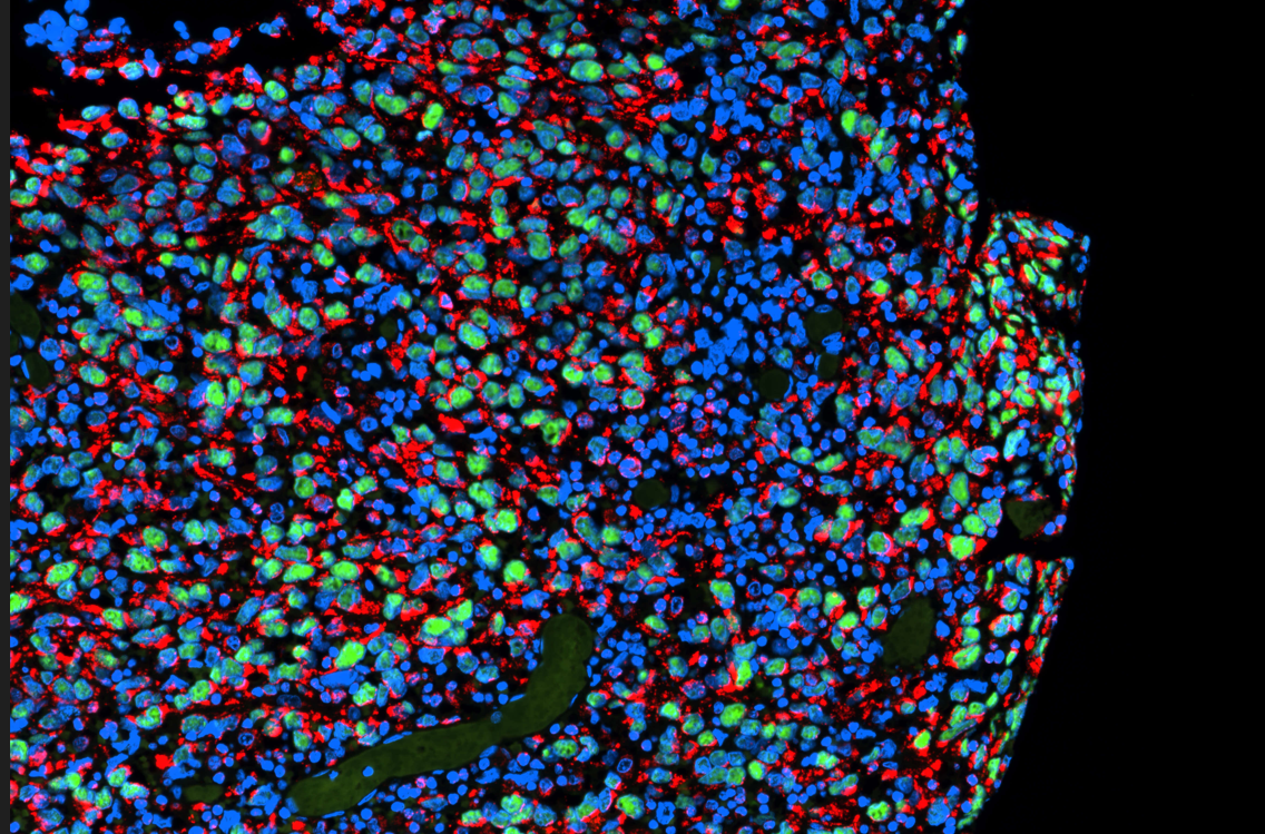
*Special stains: Masson Trichrome, Perl’s Iron, PAS, Sirius Red, Luxol F Blue, Von Kossa, Oil-O-Red, Giemsa…
Alan Spatz, MD: Core Facility Director
Naciba Benlimame, PhD: Core Facility Manager, Rm E-613, ext. 24538
Lilian Canetti: Core Facility Technician, Rm E-619, ext. 23698
Fixation is a critical step in the preparation of histological sections. If it is not carried out under optimal conditions or if fixation is delayed, a tissue specimen can be irreversibly damaged. The broad objective of tissue fixation is to preserve cells and tissue components in a “life-like state” and to do this in such a way as to allow for the preparation of thin, stained sections.
Fixation of tissues can be achieved by physical or chemical means:
In general, chemical cross-linking fixative (formaldehyde, glutaraldehyde) offers the strongest preservation of cell-tissue structures. Please keep in mind that these fixatives will often change the conformation of many antigens, and thus decrease the immunolabeling reactivity.
Notes: If you have any questions or if you are unsure about your specimen integrity, do not hesitate to contact us. We will be pleased to help you depending on your needs. Good sampling and fixation make quite a difference in obtaining reliable results.
For calcified samples, contact us before.

Ink identification on your cassettes will be cleared during xylene / alcohol processing steps!
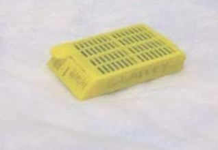
Samples should be placed in the cassettes in the same orientation that they need to be embedded (i.e., the face of the tissue that needs to be cut first should be placed towards the bottom of the cassette)
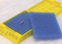
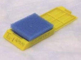
Small tissue pieces < l-2mm should be placed between blue sponges or wrapped in lens paper within a cassette.
Remember:
Formalin fixation is weak and partially reversible. Therefore, you should not store samples in buffer after formalin fixation. If you need to store samples for a long period of time, use fresh 10% formalin or 70% Ethanol and store at a cool place.
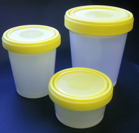
Cover all cassettes with excess 70% ETOH in a container with a screw cap, and store at 4°C until submitted to core facility. The containers may not contain more than 80%of its maximum capacity
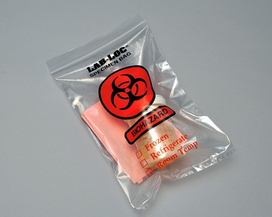
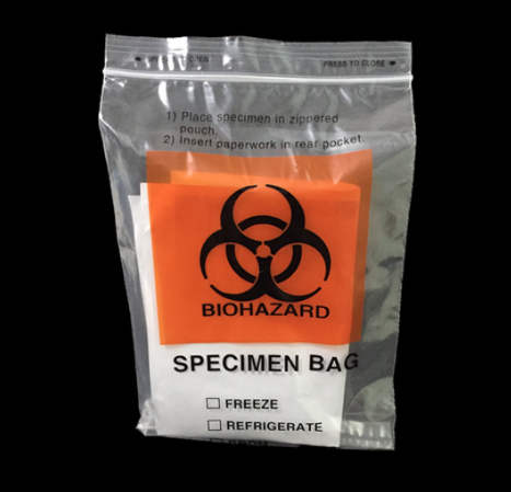
The containers must be dry from the outside and placed into plastic biohazard specimen bag. This bag must be closed properly. The closed bag with the completed histology request form can be delivered to Molecular Pathology Centre reception at E603, pavilion E.
Sample submissions that do not conform to this RPF specimen acceptance policy will be rejected.
Histology Core Personnel will contact you when samples are ready.
Remember:
All services are provided on a first-come-first-served basis.
Before bringing or sending all your samples to our facility. Make sure to fill the request form for “paraffin embedding” available on our website or contact Mrs. Lilian Canetti at ext. 23698 to get a copy. Your request will be processed more easily by including any specific detail, special needs and updated contact information (Your name, name of the PI, date and phone ext).
All those who collect, handle, send or transport specimens to the Research Pathology Facility must ensure that the container used is the appropriate one for the purpose (screw cap container that is sufficiently large volume for the number of cassettes and that fits in a biohazard bag), is properly closed, and is not externally contaminated by the contents. Sample submissions that do not conform to this RPF specimen acceptance policy will be rejected.
Before planning a frozen section project, please note that:
Frozen tissue work always needs an appointment ahead.
If tissue is not being preserved by a fixative, it is imperative that the tissue should be frozen as quickly as possible after harvesting to prevent autolysis from occurring.
Sample submissions that do not conform to this RPF specimen acceptance policy will be rejected.
Smaller size is preferred as it can be frozen faster than a larger one. But while you prepare fresh sample for frozen section, the thickness is not as critical as paraffin processing sample, it could be 1 cm or a little bit more.
Remove excess liquid surround the tissue by absorption with Kim-wipe, gauze or paper towel prior to freezing. Otherwise, these liquids will form ice crystal on the surface of tissue and prevent tissue attach to frozen embedding media (e.g., OCT compound) when the tissue is frozen embedded and cause a lot of difficulties during sectioning. This procedure only takes a few more seconds, but it will make a dramatic difference for the sectioning process later on.


Place a few drops of OCT (depends on the size of the tissue to be embedded) onto the center of the bottom of cryomold. Be careful to select the proper size embedding mold according to the size of the tissues to be embedded.
Place the unfrozen tissue sample in the OCT drop and oriented. Make sure that the side touching the bottom of the cryomold is the side you want sectioned first. Gently push the tissue with forceps to ensure that the bottom surface of the tissue is placed properly, level with the container, touching the face of the bottom and the tissue is located in the center of the mold.
Be very careful to orient the sample because it is important for the demonstration of proper morphology.
Carefully drop more OCT onto the specimen until it is completely covered. None of the tissue should remain exposed. If you are u ng a cryomold to embed your tissue, please ensure to completely fill up the mold.
Try to avoid the formation of air bubbles. Remove any bubbles inside the OCT. This is important because the air bubbles will create problems when cutting sections.
Let it settle for 15-30 seconds to allow the OCT to wet the surface of the tissue.
Place the cryomold with OCT covered sample in it on the surface of the cold isopentane/2- methylbutan or in the vapor phase right next to the liquid nitrogen with the flat side down using a long forceps.
Note: Please do not freeze the tissue by submersing it into the liquid nitrogen, it will cause the blocks to crack which makes them very difficult or impossible to section. This happens because the outside tissue begins to freeze more quickly than internal portion.
After hardening of the OCT compound (it will happen m 0.5-1 minute) wrap the OCT embedded block in foil and place it in a labeled bag or vial.
The frozen blocks can be temporarily stored in dry ice. Transfer the blocks to a liquid nitrogen storage tank (Years) or-80°C freezer (Months).
From this point over, the sample should never be thawed unless there is specific requirement.
Lady Davis Institute for Medical Research, Rm E-621
Jewish General Hospital
3755 Côte Ste-Catherine Road
Montreal, Qc
H3T 1E2
Lady Davis Institute for Medical Research, Rm E-604-1
Jewish General Hospital
3755 Côte Ste-Catherine Road
Montreal, Qc
H3T 1E2