Introduction to Flow Cytometry 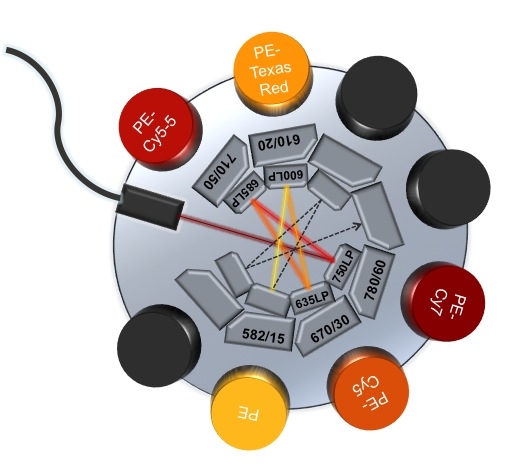
Flow
cytometry (FCM) is a versatile technology, which allows for quantification of fluorescence
and structural features of particles (most commonly cells). FCM analyzers provide
rapid quantitative analysis of particles in suspension or soluble proteins from
serum, fractioned cells, trypsinized cells or dissociated tissue. Researches
and clinicians can obtain several statistics on a single cell and population
level. Cell sorters can also analyze
particles and, in addition, can physically separate cells of interest at high
purity for downstream assays. A non-exhaustive list of different flow cytometry
assays is listed below.
There
are three major components to a flow cytometer: fluidics, optics and
electronics. With exquisite pressure control and precise flow cell or nozzle
design, the fluidics system hydrodynamicly focuses the sample and aligns the cells
in single file. The cells then flow through the heart of the system, the interrogation
point, where the fluidics meets the optics.
The
optics is composed of both light excitation and light collection modules. At
the interrogation point, lasers are used to scan each cell one after the other
to assess their physical and fluorescent parameters. The amount of light
diffracted in line with the laser (Forward Scatter; FSC) provides an indication
of size and laser diffraction at about 90o (Side Scatter, SSC)
provides an indication of cell complexity or granularity. In addition, cells
can be labeled with reporter proteins, fluorescent dyes or fluorescently
labeled antibodies, which selectively marks cells of interest. These sets of markers
or color panels must be carefully chosen to be excited by the available
excitation light source (lasers) and emit fluorescence at an emission wavelength
of light that is distinctly collected by available band-pass filters.
The
electronics components take advantage of photodiodes and ultra sensitive
photomultiplier tubes (PMTs) to convert light, defined by the band-pass
filters, into electronic pulses. These pulses are integrated, digitalized and
sent to the acquisitions station, where the data can be interpreted.
The
advantage of using FCM is that it is an extremely fast system and a relatively
small quantity of sample is needed. Furthermore, in multiparameter FCM, several
fluorescent parameters or colors are analyzed simultaneously. We can for example
identify the phenotype and ascertain viability, vitality, proliferative
capacity and cell cycle state of each cell. Therefore, since thousands of cells
can be quickly analyzed, we can identify extremely rare cell populations and
also obtain population statistics with greater accuracy.
Flow Cytometry Applications (non-exhaustive list):
Apoptosis
Autophagy
Calcium flux
Cell signaling
Cell sorting
Cell tracking
Cell viability
Cloning
Detection of soluble molecules
DNA/Cell cycle analysis
Immunophenotyping
Intracellular pH
Intracellular protein detection
Membrane potential
Phagocytosis
Proliferation
Transfection/Transduction efficiency